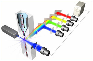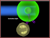
A really cool thing that a researcher can do with flow cytometry is determine the surface molecules or antigens that are present on a particular cell type. This is done by collecting a cell sample and then labeling the cells with fluorophore-labeled antibodies. You can add many antibodies (all of different colors) to look for several molecules at the same time. You can also do intracellular staining if you are looking for a particular molecule that may be found inside the cell as opposed to on its surface. Intracellular staining requires the use of a Golgi plug because you must make sure that proteins, especially proteins that you are looking for, are not shipped out of the cell by anterograde transport once you create pores in the cellular membrane to allow the fluorophores insid
 e. Once the cells have been labeled, they can be passed through the laser. The laser will excite the fluorophore to a higher energy level, the fluorophore will then return to its regular energy level causing a certain color light to be emitted, and this fluorescence will be detected by the flow cytometer. Subsequent analysis of the fluorescence intensities allows researchers to know what antigens and surface/intracellular molecules are present in a cell. This is clinically relevant because if you know what surface molecules are on a cell, you can gain insight into biological pathways or intercellular interactions of which the cells may be a part. This information can then be used to create treatments that could upregulate positive pathways or downregulate harmful pathways. As you can see, flow cytometry is extremely essential and exciting stuff. So don't forget...go with the flow.
The pictures in this post came from the website listed below. If you are interested in learning more about flow cytometry I would highly recommend this website! Más pronto...
http://www.invitrogen.com/site/us/en/home/support/Tutorials.html

No comments:
Post a Comment