Wednesday, August 25, 2010
Reflections on an Incredible Experience
I lived in the second largest city in America. I worked in one of the best neurosurgical departments at one of the best hospitals in the country. I saw multiple brain surgeries. I shadowed some of the most brilliant physicians in the world. I attended lectures, brain tumor boards, neurovascular meetings, and lab meetings all of which were focused on providing the best care possible to the patients of the Maxine Dunitz Neurosurgical Institute. I studied immunology. I gained practical research experience while working very hard in the lab. I participated in an experiment that lasted over 24 straight hours and then directly afterwards went into the OR to shadow a surgery. I saw many, many movies in the movie capital of the world. I dined, danced, and enjoyed life in LA. I visited New York and met one of my favorite movie stars. I witnessed the tremendous amount of dedication and time that is required to be a successful neurosurgeon, and I gained even more excitement and enthusiasm at the prospect of becoming one. In short, I had an absolutely amazing experience!
I feel very blessed to have participated in this program. I come away from the past three months more excited than ever to become a physician. My time at Cedars-Sinai has significantly increased my desire to become a neurosurgeon. I am more determined than ever to study hard so that I can become the best physician that I can possibly be, and I absolutely cannot wait for the day when I too can work to improve the lives of patients. I only hope that one day I will be like the neurosurgeons of Cedars-Sinai: totally committed to the well-being of their patients, doing everything possible to give their patients hope and the chance at a brighter tomorrow, and tirelessly working to cure brain cancer. I know that this summer would not have been possible without God, the members of the selection committee, my professors at Wofford, and my being a Terrier. So I would like to take this opportunity to sincerely thank every single person who had a hand in providing me with this opportunity. I will truly never forget my summer at Cedars-Sinai, and I know that it will have an immeasurable impact on my future career and life.
So how do I end this blog? Perhaps with a challenge:
Dream big.
Develop your passions.
Pour your heart into what interests you.
Spend your time on things that matter.
Go after opportunities, no matter how unattainable they may seem.
Have faith.
If you do these things, then you just might stumble upon something that defines who you are, you just might find something that you love, you just might attain your dreams.
Thanks so much for reading my blog!
-Joseph H. McAbee
Wednesday, August 4, 2010
Can we crank the bipolar up to 35, please? … microscissors … let’s get some irrigation in here!
I saw my first brain surgery a few weeks ago! It was AMAZING! It was truly awe inspiring when I stopped and thought about what all the brain controls. I mean the brain controls breathing, memory, speech, walking, personality, and pretty much everything else about us! And neurosurgeons have the responsibility of operating in this complicated control center of our bodies! They have to get in, do what needs to be done, and get out without causing neurological deficits to the patient. That is no small task. It is nerve-racking, but mostly it is really exciting. Spending time in the Cedars-Sinai OR, working with the neurosurgeons and residents, seeing brain tumor resections, watching the brain pulse along with the heartbeat, examining facial nerves, and more, have convinced me that I want to become a neurosurgeon!
Trigeminal neuralgia is a disorder characterized by intense facial pain. It typically is caused by a blood vessel putting pressure on the left or the right trigeminal nerve. The trigeminal nerve controls a multitude of functions. It transmits signals from the face to the brain, it controls jaw muscle movement, and it involves a person’s ears, eyes, nose, forehead, teeth, tongue, and more. A patient who has TN may experience intense pain in any or all of the above areas, typically on one side of the face, many times throughout the day. Attacks can be triggered by something as simple as brushing one's teeth or a cool breeze. It is a debilitating disease that gets worse over time. A neurosurgeon can correct the problem through microvascular decompression. He/she can make an incision behind the ear, cut open the skull, move the brain to expose the trigeminal nerve, separate the nerve from the blood vessel, and place a Teflon pad between the nerve and vessel to insure that the blood vessel can no longer put pressure on the nerve. The patient should recover well with little to no pain or facial numbness.
Tuesday, July 20, 2010
Tuesday, July 13, 2010
Bregma marks the spot. Well, sort of.
I apologize that this post is so long in coming. I typed this post up last week, but unfortunately I accidentally erased it all. Smooth, I know. I did not have time to retype it this weekend, as my parents were visiting me in LA. We had a lot of fun! We did many of the tourist activities in LA which was cool since I hadn’t done many of those things yet even though I have been living in LA for two months! I will post those pictures soon.
Several weeks ago, I was able to assist in my first surgery ever! It was awesome. Before I begin my description on tumor implantation, I want to reiterate that all procedures are done to ensure that the animals are treated in the most humane way possible. The mice are first anesthetized. Then their heads are shaved. Once they are completely under anesthesia, they are placed on the stereotactic frame, as shown above. After the mice are secure, betadine is applied to the incision area and the skin covering the skull is cut. The skull is exposed and the bregma is located. The bregma is actually easier to locate when the skull dries slightly. The bregma is the point of intersection between the coronal suture (horizontal line in the picture to the left) and the sagittal suture (vertical line) where the frontal and parietal bones meet. We use the bregma as the “zero point” or the point of reference. Once we find the bregma, we utilize the stereotactic frame to find the correct point in the right hemisphere where we will inject the tumor cells. A burr hole is created in the skull using a hand drill, a needle is inserted into the hole, and 50 K tumor cells are injected into the hole. After injection, bone wax is applied to cover the burr hole, the skin is clipped together, and pain killers are injected subcutaneously. The mice are observed closely for several hours after surgery and then each day until terminal symptoms become apparent. This particular type of surgery can be performed to derive tumor lines or to observe the effects that different treatment types have on tumor growth and mouse survival time.
Wednesday, July 7, 2010
To Perfuse or Not to Perfuse... That's Not Even a Question!
I would like to preface my discussion of perfusions by saying that all work that we do on mice is done following a set protocol that has been created in collaboration with veterinarians, research scientists, and healthcare professionals to ensure the most humane methods of euthanasia. Also, all mice studies are only done when other routes of experimentation are unavailable and there is sufficient need for the experiment.
So why is doing a perfusion the definite route to go when you are seeking to resect a brain or spleen for flow cytometry or other types of analyses? I am glad you asked. It is because perfusions allow a scientist to remove the blood from the organ of interest which cuts down on auto fluorescence by RBCs. First, the mouse is anesthetized with a ketamine + xylazine IP injection. The mouse must be completely unconscious before the procedure can begin so that it will not feel any pain, and this can be tested by squeezing its paws and observing the reaction. Next, a dissection is performed to expose the abdominal and thoracic cavities on the ventral side of the mouse. The ribs are removed and the heart is exposed. After exposing the heart, the aorta is cut and a small hole is cut in the left ventricle. A butterfly needle is placed into the left ventricle and PBS is injected into the mouse. The mouse’s heart will carry the PBS throughout the body which will rid the body of RBCs and cause the organs to turn white in color. If the brain tissue needs to be fixed, then PFA can also be injected through the left ventricle to do so. After this procedure is done, the required organs can be removed and properly stored for analysis.
Another important procedure for researchers that are studying cancer is tumor implantation. Tumor implantation is two things: 1. It is a very neat procedure, and 2. It is the topic for my next blog… Stay tuned.
Tuesday, June 29, 2010
And The #1 Most Important Technique in Scientific Research is......
Saturday, June 19, 2010
Living the L.A. Life
I went to Santa Monica today. I hung out on the pier for a little while, which is equipped with its own roller coaster, Ferris wheel, and food court, and then I went on the beach to read, listen to music, and admire the Pacific Ocean. I had a lot of fun, and luckily I did not get too sunburned! When I got back to my apartment, I walked down to San Vincente and got some supper at a small cafe in downtown Brentwood. Last weekend, I found a neat outdoor mall just outside of Beverly Hills with the nicest movie theater ever. This was a big deal to me because I am a huge movie buff (something that I have inherited from my father) and so I plan to spend a lot of time there this summer. It had three stories and had assigned seating! I saw Prince of Persia. It was great! I am not a shopper, but if I were it would be easy for me to do so since I drive right past Rodeo Drive on my way to work each day.
There is never a shortage of things to do in L.A. However, in addition to working full-time during the week and having fun on the weekends, I have to find time to cook, clean, wash clothes, iron, go to the grocery store, exercise, and study. It is a little different from being at Wofford where my meals are made for me each day and I can get my clothes washed every time I go home to visit. I like it though. I am truly living in the "real world," and it is great practice for the future.
One thing that I really like about L.A. is the interesting and eclectic group of people that I interact with on a daily basis. At the lab I am working in I get the opportunity to work closely with people of many different races and backgrounds. Each person has his/her own ideas, beliefs, and languages. I have enjoyed getting to know my labmates and many other people at Cedars-Sinai. L.A. is really teaching me that we truly do live in an internationally oriented world, and that advances in science, economics, and politics are made much easier when all people work together in a spirit of cooperation and brotherhood.
Even though I am a Woodruff boy and I am used to the slower pace of Spartanburg, South Carolina, I really love L.A. Sure it is busy, crowded, and crazy at times, but it is also beautiful, fun, and an exciting place to live and work. Oh and by the way...GO LAKERS!!!!!!!!!! NBA CHAMPS!!!!!!!!!!
Thursday, June 17, 2010
Go with the Flow
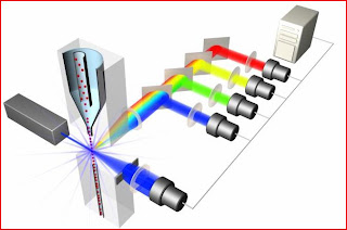
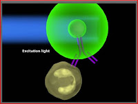
A really cool thing that a researcher can do with flow cytometry is determine the surface molecules or antigens that are present on a particular cell type. This is done by collecting a cell sample and then labeling the cells with fluorophore-labeled antibodies. You can add many antibodies (all of different colors) to look for several molecules at the same time. You can also do intracellular staining if you are looking for a particular molecule that may be found inside the cell as opposed to on its surface. Intracellular staining requires the use of a Golgi plug because you must make sure that proteins, especially proteins that you are looking for, are not shipped out of the cell by anterograde transport once you create pores in the cellular membrane to allow the fluorophores inside. Once the cells have been labeled, they can be passed through the laser. The laser will excite the fluorophore to a higher energy level, the fluorophore will then return to its regular energy level causing a certain color light to be emitted, and this fluorescence will be detected by the flow cytometer. Subsequent analysis of the fluorescence intensities allows researchers to know what antigens and surface/intracellular molecules are present in a cell. This is clinically relevant because if you know what surface molecules are on a cell, you can gain insight into biological pathways or intercellular interactions of which the cells may be a part. This information can then be used to create treatments that could upregulate positive pathways or downregulate harmful pathways. As you can see, flow cytometry is extremely essential and exciting stuff. So don't forget...go with the flow.
The pictures in this post came from the website listed below. If you are interested in learning more about flow cytometry I would highly recommend this website! Más pronto...
http://www.invitrogen.com/site/us/en/home/support/Tutorials.html